Inner Awakening and cellular Transformation
This text was machine-transcribed from the printed book and may contain errors. Select the words and choose “Report a mistake”.
THE SCIENCE OF MITOCHONDRIAL FUNCTION
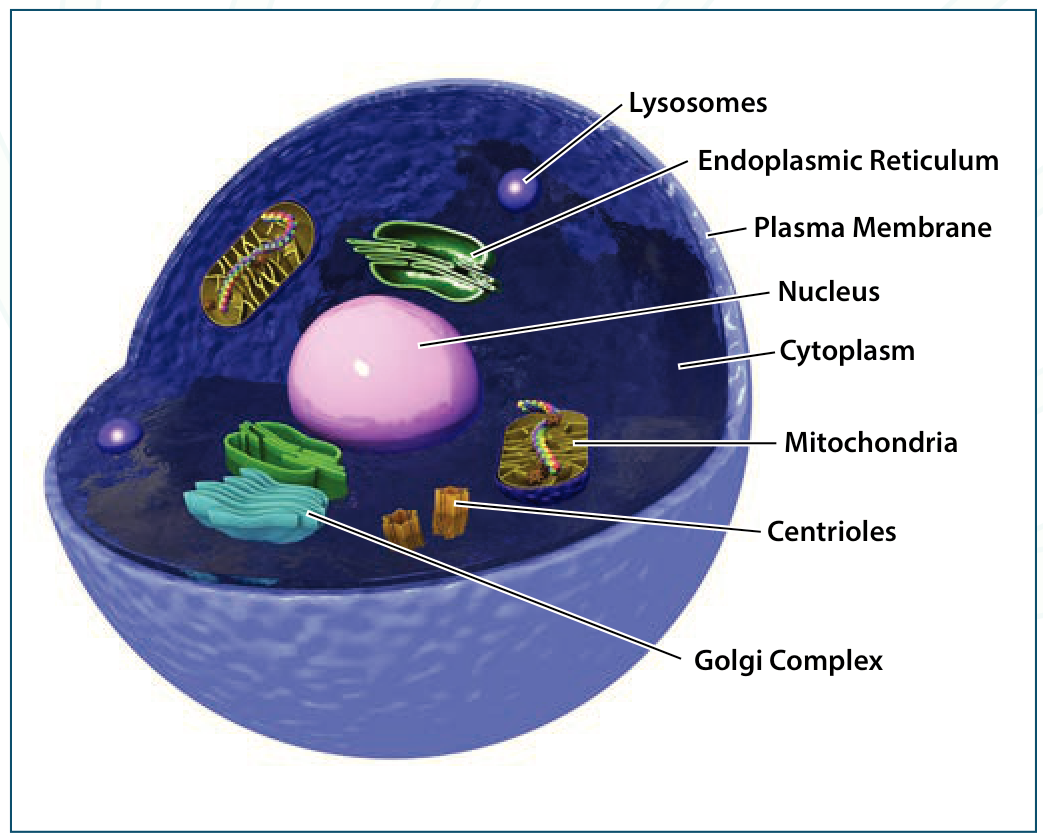
The human body is essentially an assembly of 50 trillion individual cells living in unison, working towards a common goal. Each cell is by itself a complete living entity with its own processes for sustenance of life, reproduction and so on. Most of the components of the cells are common across the board fulfilling basic common needs, yet each have their own specialized departmental work, be it secreting hormones, enzymes and so on. All these cells are powered by a single battery within the cell called the mitochondrion.
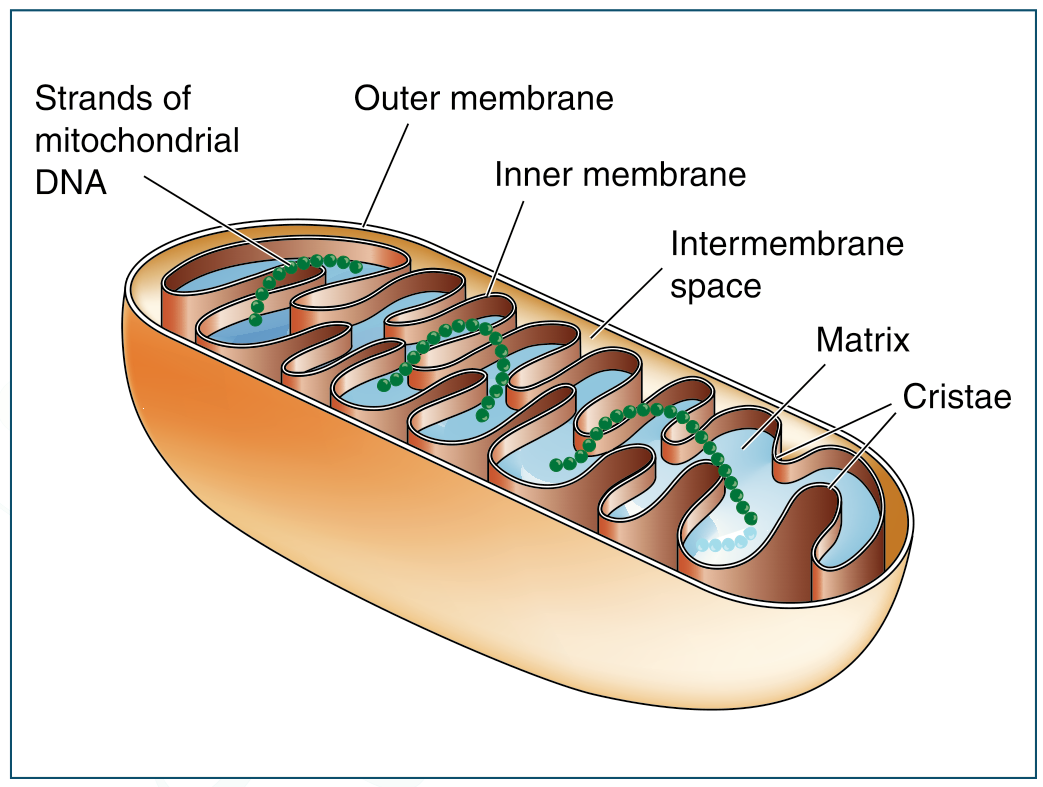
The mitochondrion is an interesting structure, only 1 micron in length, situated outside the nucleus in the cytoplasm of the cell. Mitochondria are the cell's power producers. They convert energy into forms that are usable by the cell. They are the sites of cellular respiration which ultimately generate fuel for the cell's activities. Their function is to optimize the utilization of energy producing substances like glucose, and to produce ATP which is the energy molecule of the cell. Without mitochondria, the cell can produce only 2 ATPs per glucose
The mitochondria have their own set of DNA, which allows them to function independently. Unlike the DNA in the nucleus of the cell, which is covered by a protective layer of histones, the mitochondrial DNA is naked and thus exposed to increased possibilities of damage. As it is, the repair mechanism of the mitochondrial DNA is not as efficient as that of the nuclear DNA. As we age, significant errors creep into the mitochondrial DNA which go unrepaired or inappropriately repaired. This leads to malfunction in the electron transfer chain of the mitochondria. Instead of producing pure energy in the form of ATP, a non-productive oxygen species called reactive oxygen species (ROS) is produced. This ROS damages cellular structures including the exposed mitochondrial DNA, leading to a vicious cycle of dysfunction and aging, which in turn leads to cell death.
molecule. The mitochondrion, on the other hand, can produce 32 ATPs per molecule of glucose, which is a 1700% in efficiency. Mitochondria are also involved in other cell processes such as cell division and growth, as well as cell death.
MITOCHONDRIAL ASSAY—
THE STUDY
a. 10 random blood samples from Inner
Awakening (IA) participants from
above 50 years of age as study group
b. Blood samples were taken on Day 0
and Day 18
c. Labeling was double blinded and sent
to the laboratory for MTT assay
d. All participants received energy
darshan from Nithyananda in addition
to 21 days of yoga, meditation, exercise
and other processes.
LABORATORY ANALYSIS
The MTT assays were performed at Post
Graduate Department of Biotechnology,
Mangalore University.
All results including the physical, physiological measurements and the MTT assay were statistically evaluated by professional biostatistician Dr. K. P. Suresh, Scientist (Bio Statistics), National Institute of Animal Nutrition and Physiology, Bangalore.
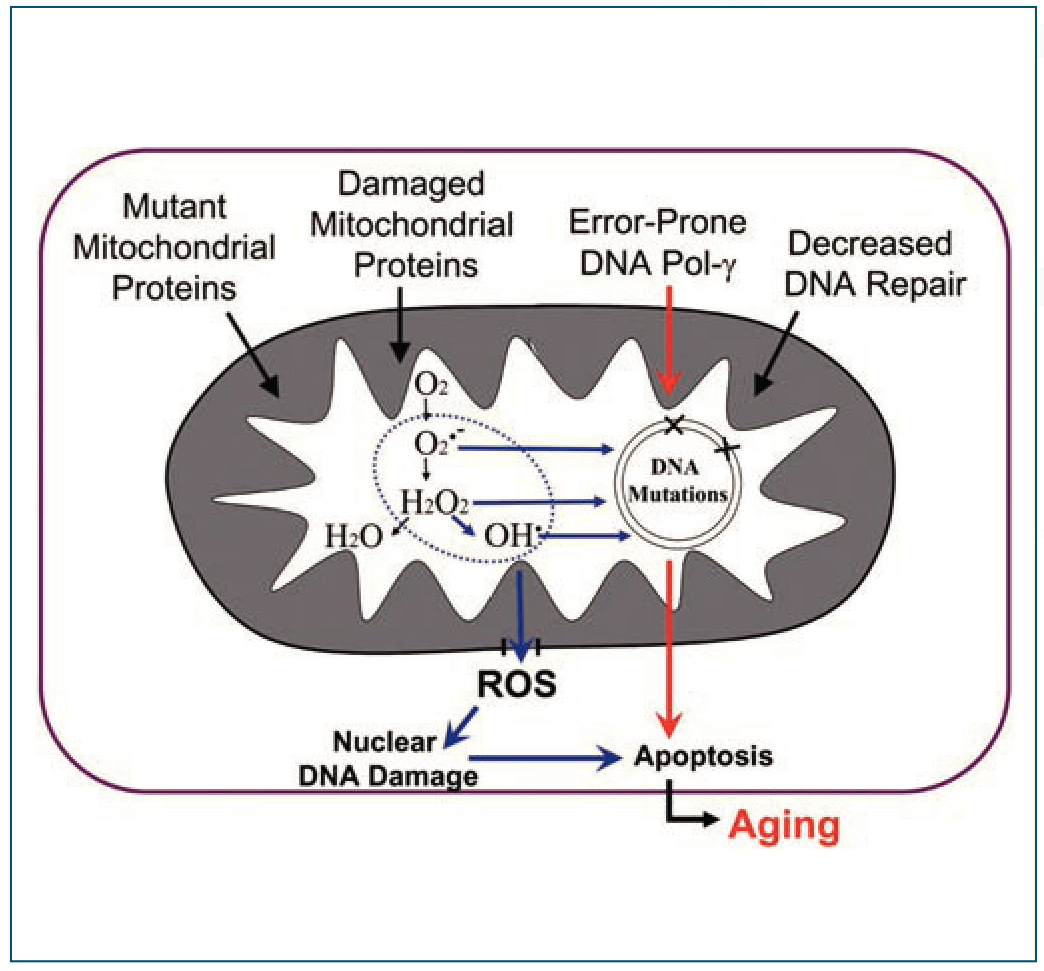
Fig: ROS mediated cell damage Since the etiology of many diseases is currently being quoted as related to free-radical mediated damage, mitochondria seemed to be the right subject for study and that is where we started. Genetics also plays a major role in the etio-pathogenesis of diseases. Genetic studies were carried out on participants who had significant health recoveries. QEEG studies were also done to study the effect on brain waves.
MATERIAL AND METHODS:
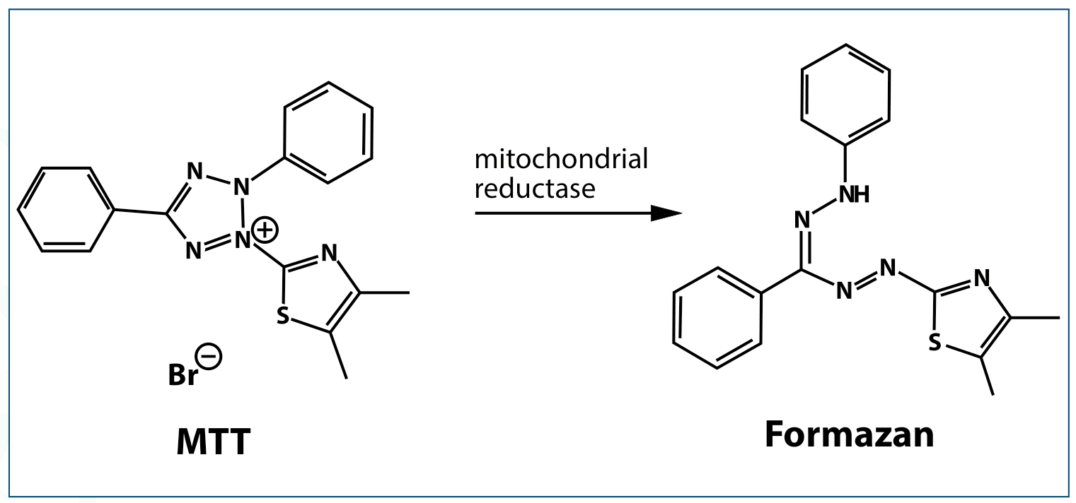
This colorimetric assay is dependent on the ability of viable cells to metabolize a water-soluble tertazolium salt (yellow color) into a water insoluble formazan product (purple color). It measures the activity of cellular respiratory enzymes that reduce MTT. Since only viable cells perform this activity, the assay is used to measure the viability and proliferation of cells.
PROCEDURE: ISOLATION OF MONONUCLEAR CELLS (MNC)
Human Mononuclear cells can be separated from heparinized blood samples or from the buffy coat of centrifuged blood sample with sodium citrate as an anticoagulant after removal of the erythrocytes and the plasma. The residual fraction (25–40 ml) contains more than 90% of the leukocytes, 10% of the erythrocytes, and 5% of the plasma.
- Dilute buffy coats 1:4 (v/v) or heparinized blood samples 1:2 (v/v) with Dulbecco’s Phosphate buffered saline (PBS) without Ca²⁺ and Mg²⁺.
- To a 10 ml centrifuge tube add 2 ml of defibrinated or anticoagulant treated blood and an equal volume of balanced salt solution (final volume 4 ml).
- Mix the blood and buffer by inverting the tube several times or by drawing the mixture in and out of a pipette.
MTT is (3-(4, 5 Dimethylthiazol-2-yl)-2, 5-di phenyltetrazolium bromide
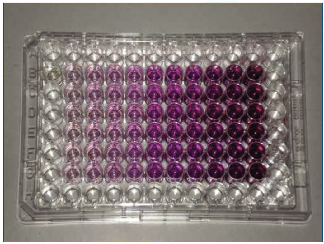
A Micro-titer Plate after MTT Assay
Tetrazolium salts are converted by metabolically active cells to formazan, which can be quantified by measuring absorbance at 570 nm.
Procedure for isolation of mononuclear cells
- Invert the Ficoll-Paque media bottle several times to ensure thorough mixing.
- Using aseptic techniques, add Ficoll-Paque media (3 ml) to the centrifuge tube.
- Carefully layer the diluted blood sample (4 ml) onto the Ficoll-Paque media solution.
- Centrifuge at 400 g for 30 to 40 minutes at 18°C to 20°C (brake should be turned off).
- Draw off the upper layer containing plasma and platelets using a sterile pipette, leaving the mononuclear cell layer undisturbed at the interface.
- Transfer the layer of mononuclear cells to a sterile centrifuge tube using a sterile pipette.
Washing the cell isolate
- Estimate the volume of the transferred mononuclear cells. Add at least 3 volumes (~ 6 ml) of balanced salt solution to the mononuclear cells in the centrifuge tube.
- Suspend the cells by gently drawing them in and out of a pipette.
- Centrifuge at 400 to 500 × g for 10 to 15 minutes at 18°C to 20°C.
- Remove the supernatant.
- Re-suspend the mononuclear cells in 6 to 8 ml balanced salt solution. 6. Centrifuge at 400 to 500 × g (or 60 to 100 × g for removal of platelets) for 10 min at 18°C to 20°C.
7. Remove the supernatant.
8. For cell counts, dilute first 1:10 in
supplemented RPMI -1640, and then
1:2 with 0.1% trypan blue solution, and
count the cells with a Hemocytometer.
MTT Assay—Steps:
1. Remove the culture medium from cell monolayers cultured in 96-well plates.
2. Reconstitute each well with 0.2 mL
supplemented RPMI-1640 containing
1 mg/mL MTT.
3. Incubate the cultures for 2–4 hours at 37°C.
4. Remove the supernatants from the wells.
5. Add 0.2 mL per well of a lysing reagent containing 0.04 N HCl in isopropanol.
6. Mix the content of the wells thoroughly.
7. Read the plates in an automated
microplate spectrophotometer at 570
and 630 nm as reference
References: Mossman, T. (1983). Rapid colorimetric assay for cellular growth and survival. Journal of Immunological Methods, 65(1-2), 55-63. Sieuweters, A. M., Klijn, J. G., Peters, H. A., & Foekens, J. A. (1995; Nov. 33). MTT tetrazolium salt assay scrutinized. Eur J Clin Chem Clin Biochem, 11, 813-823. Darzynkiewicz, Z., Li, X., & Gong, J. (1994). Assays of cell viability: discrimination of cells dying by apoptosis, Methods in Cell Biology, 41, 15-38.
Increase in
MITOCHONDRIAL ACTIVITY During inner Awakening
| MTT Assay | ||||||
|---|---|---|---|---|---|---|
| Sample No | Day 0 | Day 20 | ||||
| Reading 1 | Reading 2 | Average | Reading 1 | Reading 2 | Average | |
| 1 | 0.28 | 0.148 | 0.214 | 1.787 | 1.316 | 1.5515 |
| 2 | 0.242 | 0.109 | 0.1755 | 1.567 | 0.747 | 1.157 |
| 3 | 0.041 | 0.211 | 0.126 | 1.864 | 0.952 | 1.408 |
| 4 | 0.121 | 0.123 | 0.122 | 1.942 | 0.728 | 1.335 |
| 5 | 0.021 | 0.157 | 0.068 | 1.521 | 0.796 | 1.1585 |
| 6 | 0.04 | 0.156 | 0.098 | 1.495 | 0.722 | 1.1085 |
| 7 | 0.007 | 0.308 | 0.1575 | 1.533 | 0.763 | 1.148 |
| 8 | 0.011 | 0.363 | 0.187 | 1.177 | 0.468 | 0.8225 |
| 9 | 0.019 | 0.019 | 0.019 | 1.486 | 1.064 | 1.275 |
| 10 | 0.054 | 0.099 | 0.0765 | 0.553 | 0.658 | 0.6055 |
| Average | 0.1 2435 | 1.15695 |
STATISTICAL ANALYSIS
Null Hypothesis: There is no significant difference in the mean value between two groups, i.e. μ1 = μ2 Alternate Hypothesis: There is a significant difference in the mean value between two groups, i.e. μ1 ≠ μ2 Level of Significance: α = 0.05
Statistical test used: t test Decision: The p value and the level of significance are compared. If p < 0.05, the null hypothesis is rejected and the alternate hypothesis is accepted. If p ≥ 0.05, the null hypothesis is accepted. Computations: The tables below give us the various computations and the p value.
Comparison of the change in MTT Assay at Day 0 and Day 20 in study group:
| Group | Day | Mean | Std Dev | Mean Difference | t | p-value |
|---|---|---|---|---|---|---|
| Study Group | Day 0 | 0.12 | 0.06 | -1.033 | -12.084 | <0.001 |
| Day 20 | 1.16 | 0.28 |
*Denotes significant difference
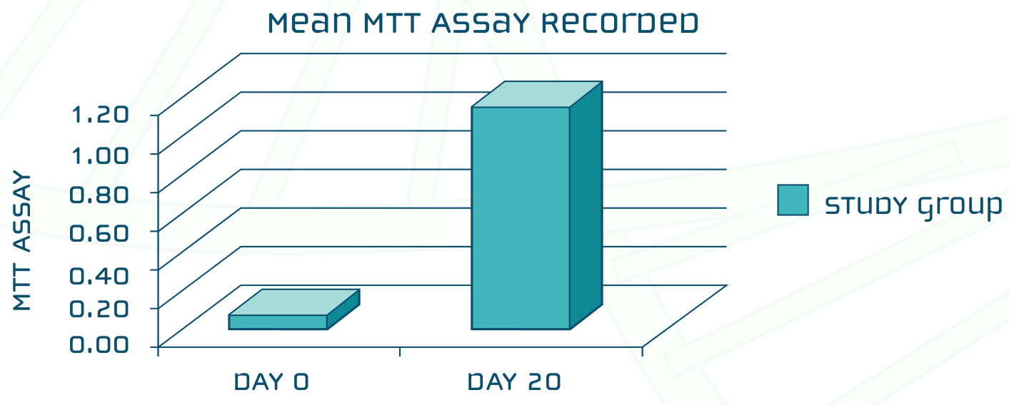
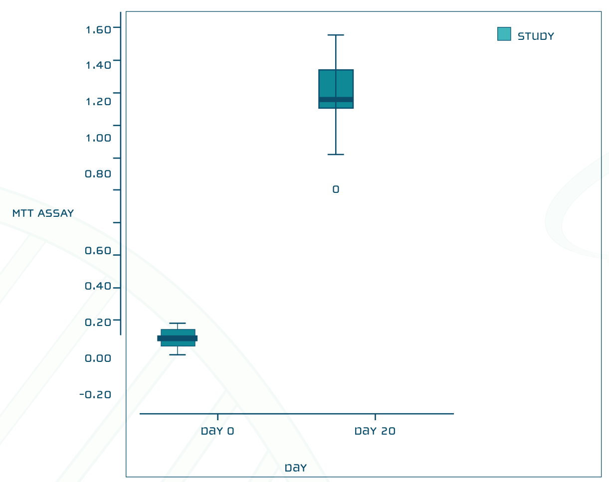
In study group, there was an increase in mean MTT Assay from Day 0 to Day 20. The increase in MTT Assay from Day 0 to Day 20 in Study Group was found to be statistically significant (p < 0.001).
Summary:
• The MTT assay is a well-established
protocol for the study of cell viability
and metabolism (Mitochondria).
• There was a dramatic increase in mitochondrial activity in the study group
• Normally, processes like yoga or exercise or meditation increase mitochondrial activity by about 40-50%.
• This study conclusively showed approximately 1000% increase of mitochondrial activity (cell viability) in the IA participants.
Conclusion:
• Energy darshan leads to Kundalini
awakening to a statistically significant
degree
• The addition of yoga, meditation and
exercise along with energy darshan leads
to greater increase in cellular energy