Neurological Research at Apollo Hospital
This text was machine-transcribed from the printed book and may contain errors. Select the words and choose “Report a mistake”.
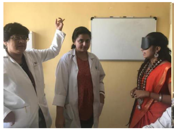
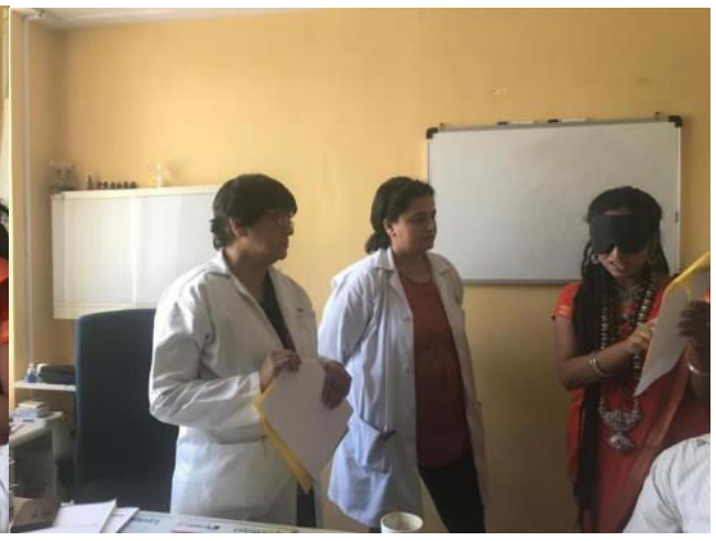
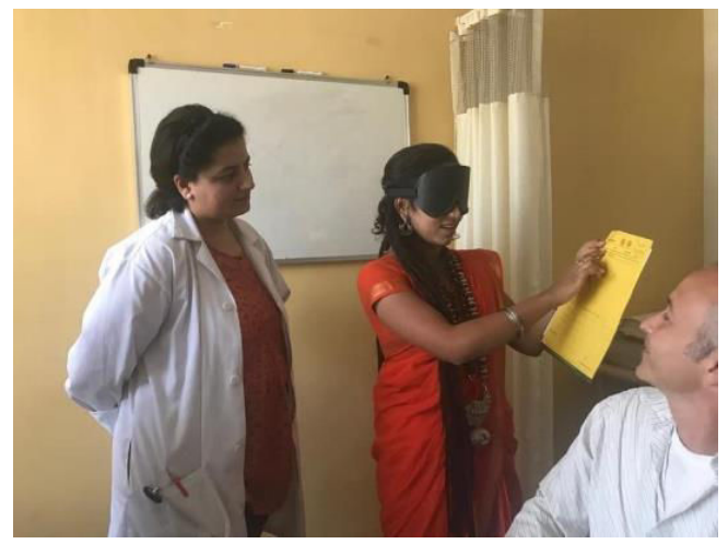
In Spring 2019, neurological research was conducted in Apollo Hospital, Ahmedabad, India under the supervision of Dr. Peter Bloomfeld, faculty of the University of California, Los Angeles. Three tests were conducted: VEP EEG.
TEST 1: Seeing without Activity in Visual Tract
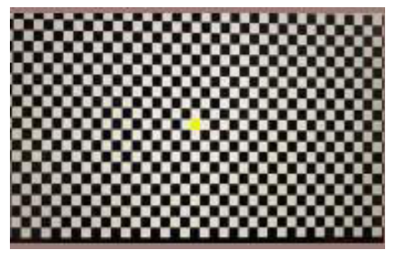
Checkerboard used for testing
Controls:
5 Balasanths (students of Nithyananda Gurukul) and 5 children (same age)
Tests Conducted:
Visual Evoked Potential (VEP)
Hypothesis:
See Appendix for detailed studies
Third Eye vision requires activity in the visual tract.
Study:
Measure neuronal activity along the normal optic pathways, in which visual data is received in the retina and travels through the optic tracts to the occipital lobe of the brain for processing.
Protocol - VEP Testing
1. Subject undergoes normal VEP testing of each eye to demonstrate normal visual pathways are intact.
2. Subject has both eyes taped shut & acrylic blackout blindfold is applied on top.
3. Subject undergoes VEP testing w/ blindfold in place while subject tries to focus on the checkerboard.
4. During testing subject is asked randomly at 10 times whether the red dot is covered or not; the # of times correctly identified is documented.
5. During testing 5 different objects are identified into the field of VEP testing; the subject is asked to identify the object. The number of times the object correctly identified is documented.
6. After testing, without any adjustment to blindfold, 9 words are introduced from the InspireMe random word generator. The number of words the subject can successfully read is documented.
7. Baseline is obtained on all subjects where they are instructed to try not to focus on the checkerboard or see through the blindfold.
Results:
| Subject | Scenario | Vision | EEG measurement |
|---|---|---|---|
| Balasanth | Baseline - Eyes open | YES | Normal brain activity |
| Children | Baseline - Eyes open | YES | Normal brain activity |
| Balasanth | Mindfold blindfold Eye Patch | YES (ZERO THOUGHTS EXPERIENCED) | • Alpha waves (8-12Hz) which commonly happen when people have their eyes closed. • Very distinct high amplitude high beta jigsaw pattern, range of 22-25 Hz |
| Children | Mindfold blindfold Eye Patch | NO | Alpha waves (8-12Hz) which commonly happen when people have their eyes closed. |
TEST 2: Super Human Beta Activity
Controls:
5 Balasanths (students of Nithyananda Gurukul) and 5 children (same age)
Tests Conducted:
ElectroEncephaloGram (EEG)
Hypothesis:
Third Eye awakening activity has no change in brain wave activity Study: Measure brain wave activity throughout the brain while performing various activities.
Protocol - EEG Testing
1. Subject undergoes normal baseline Video EEG w/ normal set up.
2. Minimum of one-minute baseline data is recorded.
3. Minimum of one minute where subject seeks to go into a space of oneness is recorded.
4. Subject is blindfolded.
5. Minimum of one minute where the subject is asked to read blindfolded is recorded.
6. During this time, 9 words are introduced from the InspireMe random word generator. The number of words the subject can successfully read is documented.
Results:
| Subject | Scenario | Vision | EEG measurement |
|---|---|---|---|
| Balasanth | Baseline - Eyes open | YES | Normal brain activity |
| Children | Baseline - Eyes open | YES | Normal brain activity |
| Balasanth | Mindfold blindfold Eye Patch | YES (ZERO THOUGHTS EXPERIENCED) | • Alpha waves (8-12Hz) which commonly happen when people have their eyes closed. • Very distinct high amplitude high beta jigsaw pattern, range of 22-25 Hz |
| Children | Mindfold blindfold Eye Patch | NO | Alpha waves (8-12Hz) which commonly happen when people have their eyes closed. |
TEST 3: Minimal Brain Activity
Controls:
5 Balasanths (students of Nithyananda Gurukul) and 5 children (same age)
Hypothesis:
Third Eye awakening activity has no change in brain wave activity
Study:
Measure brain activity in various parts of the brain
Method:
Functional magnetic resonance imaging (fMRI)
Protocol: fMRI
1. With foam & acrylic blindfold applied, the subject is exposed to 5 colored lights in a different sequence.
2. With a blindfold in place, the subject is then showed pictures and/or words.
3. After each round of fMRI recording is completed, the number of lights in sequence and pictures and/or words the subject could identify while blindfolded is documented.
4. Subject undergoes normal baseline fMRI.
5. With eyes open, 5 different colored lights flashed for the subject.
6. With eyes open, 5 different colored lights flashed for the subject.
7. With eyes open, the subject is showed with pictures.
Results:
| Subject | Scenario | Vision | fMRI measurement |
|---|---|---|---|
| Balasanth | Baseline - Eyes open, images | YES | Normal brain activity |
| Children | Baseline - Eyes open, images | YES | Normal brain activity |
| Balasanth | Mindfold blindfold - images | YES | very little brain activity |
| Children | Mindfold blindfold - images | NO | midbrain and occipital area (sign of anxiety) |
| Balasanth | Mindfold blindfold - images, lights flashing | YES | very little brain activity |
| Children | Mindfold blindfold - images, lights flashing | NO | midbrain and occipital area (sign of anxiety) |