Conclusions
This text was machine-transcribed from the printed book and may contain errors. Select the words and choose “Report a mistake”.
The brain is the body’s most complex organ, containing more than 100 billion neurons, each of them in chemical and electrical conversation with up to 10,000 other neurons. Its sheer capacity to process information is astonishing.
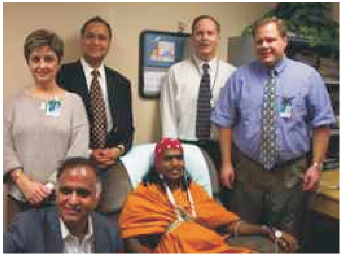
Remarkably, that complexity presents little difficulty for Swami in managing his brain activity. Swami’s mind – his thoughts, emotions and intellect – control his brain. He can, in a very fluid, easy way, shift his brain function and alter his brainwaves.
More than answering questions, the voyage we took into the mind of a mystic brings intriguing questions for study:
Are there techniques we can learn and teach that will bring balance and peace into people’s lives? Can we invoke a healing response or accelerate healing through specific training? Can we learn techniques that will allow us to control pain or alter the course of a disease? Can we learn to activate what I call our D-spot, thus putting us in instant connection to delight or the divine?
The results from our study of Swami are new pages in our world’s growing book of research on the brain. There continue to be indications that the human mind may be able to choose to heal the body. We’re now looking at the possibility of people learning and acquiring these healing capabilities, an event of immense benefit for humankind. The potential for altering the rates and progression of many diseases – heart disease, cancer, arthritis, alcoholism and many others – is beginning to look achievable.
Swami is a bridge between the invisible, ancient world of mysticism and the modern, visible world of science and discovery. As brain research continues on a widespread basis, and as we appropriately bring the phenomena of mysticism into the realm of science for further study, we are taking strides on a path of hope and health.
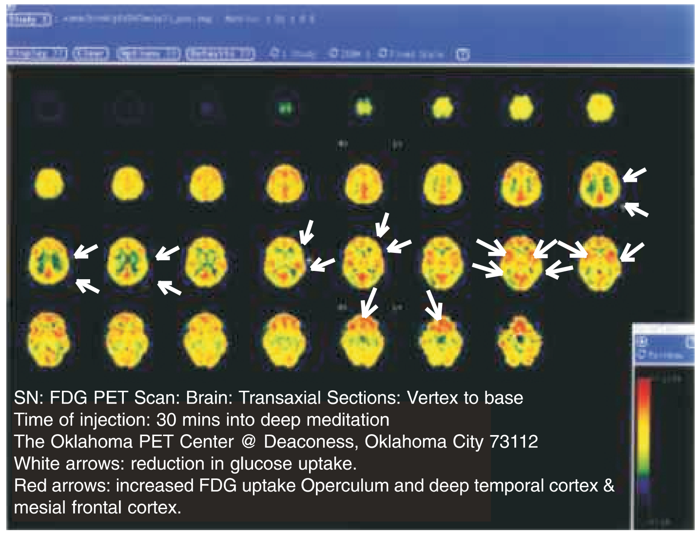
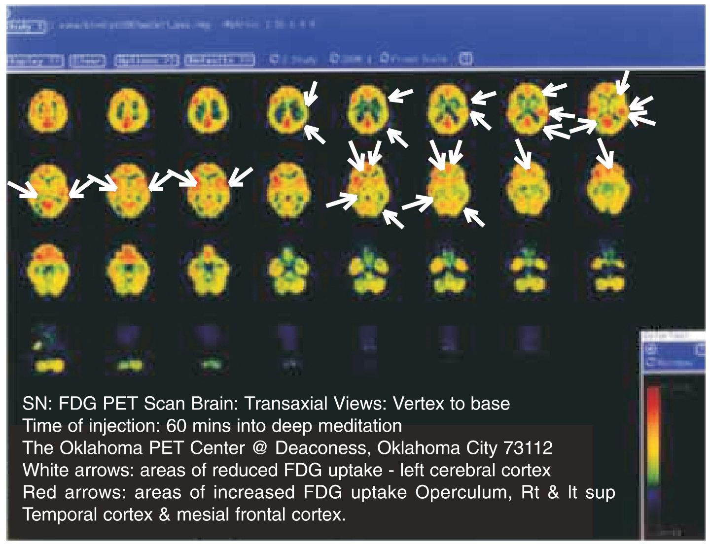
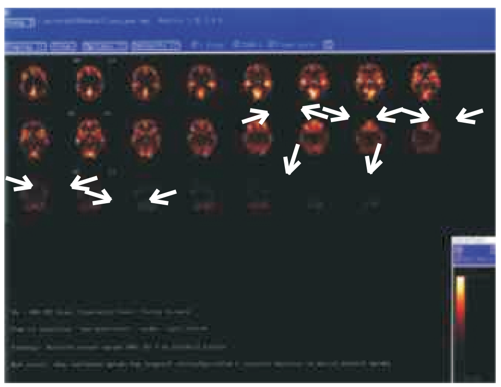
Following are images from the PET SCAN:
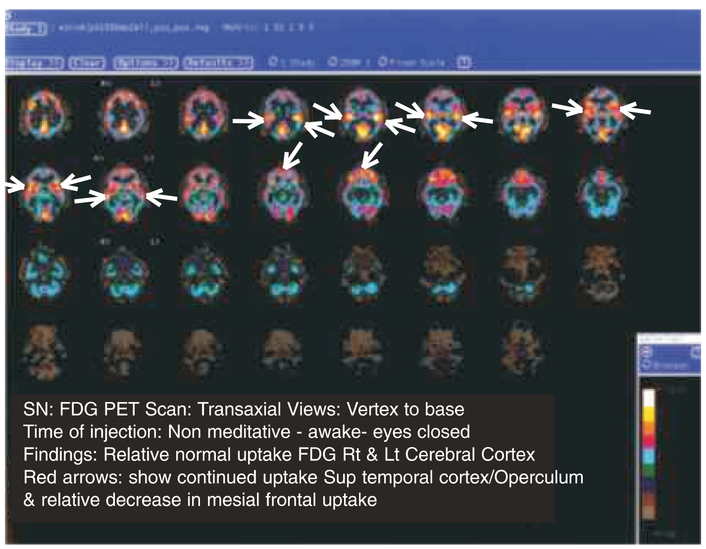
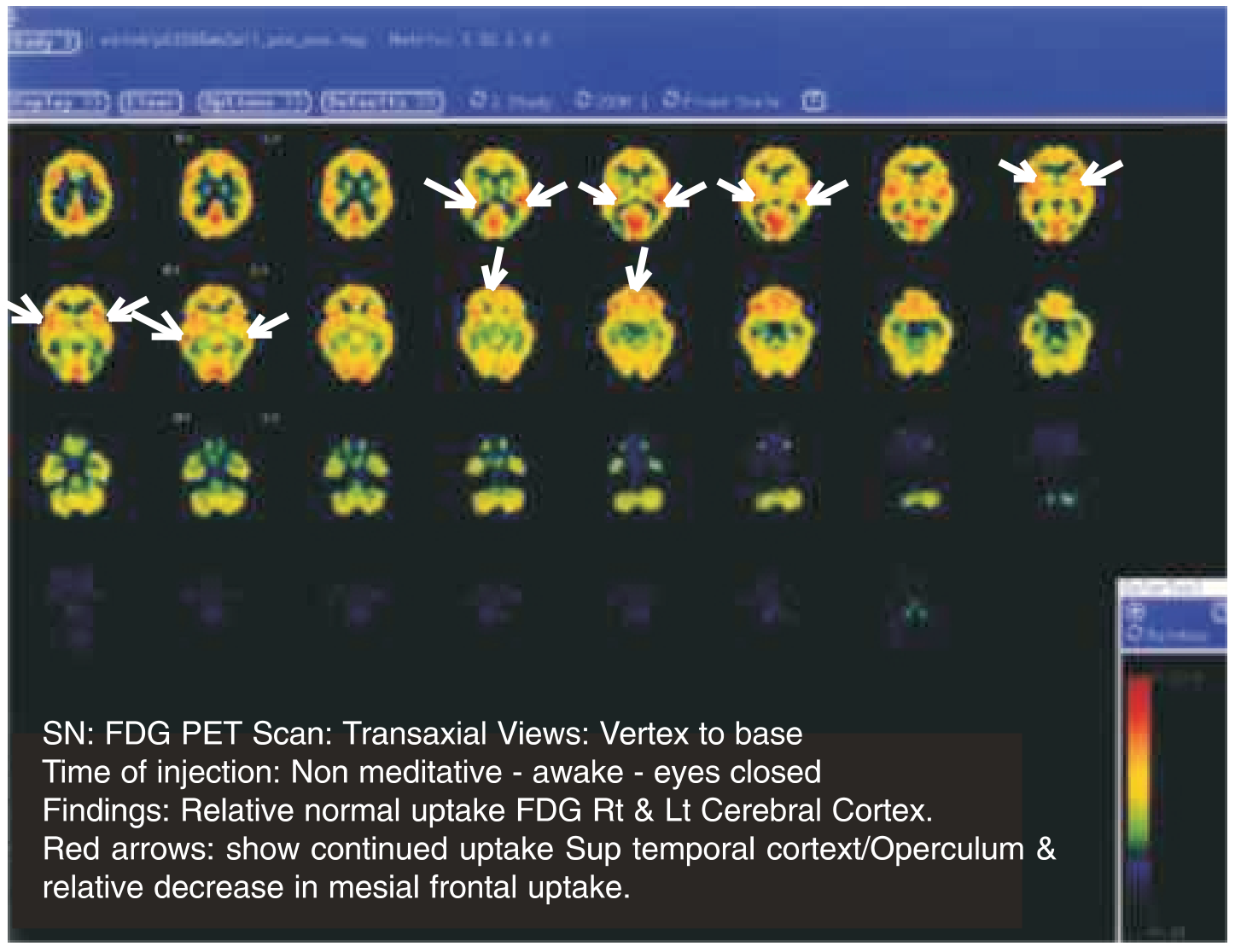
Time of injection: Non meditative - awake - eyes closed Findings: Relative normal uptake FDG Rt & Lt Cerebral Cortex. Red arrows: show continued uptake Sup temporal cortext/Operculum & relative decrease in mesial frontal uptake.
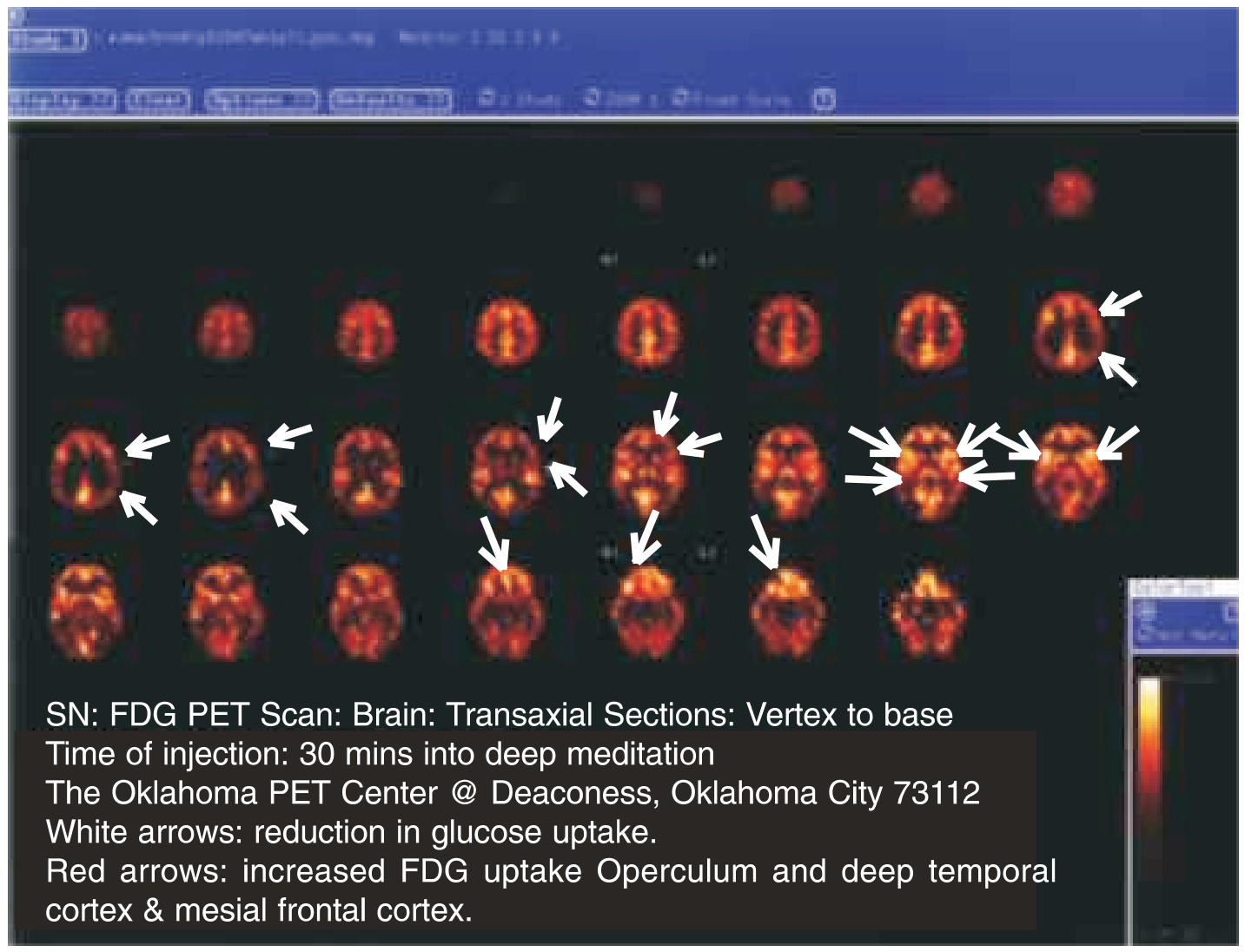
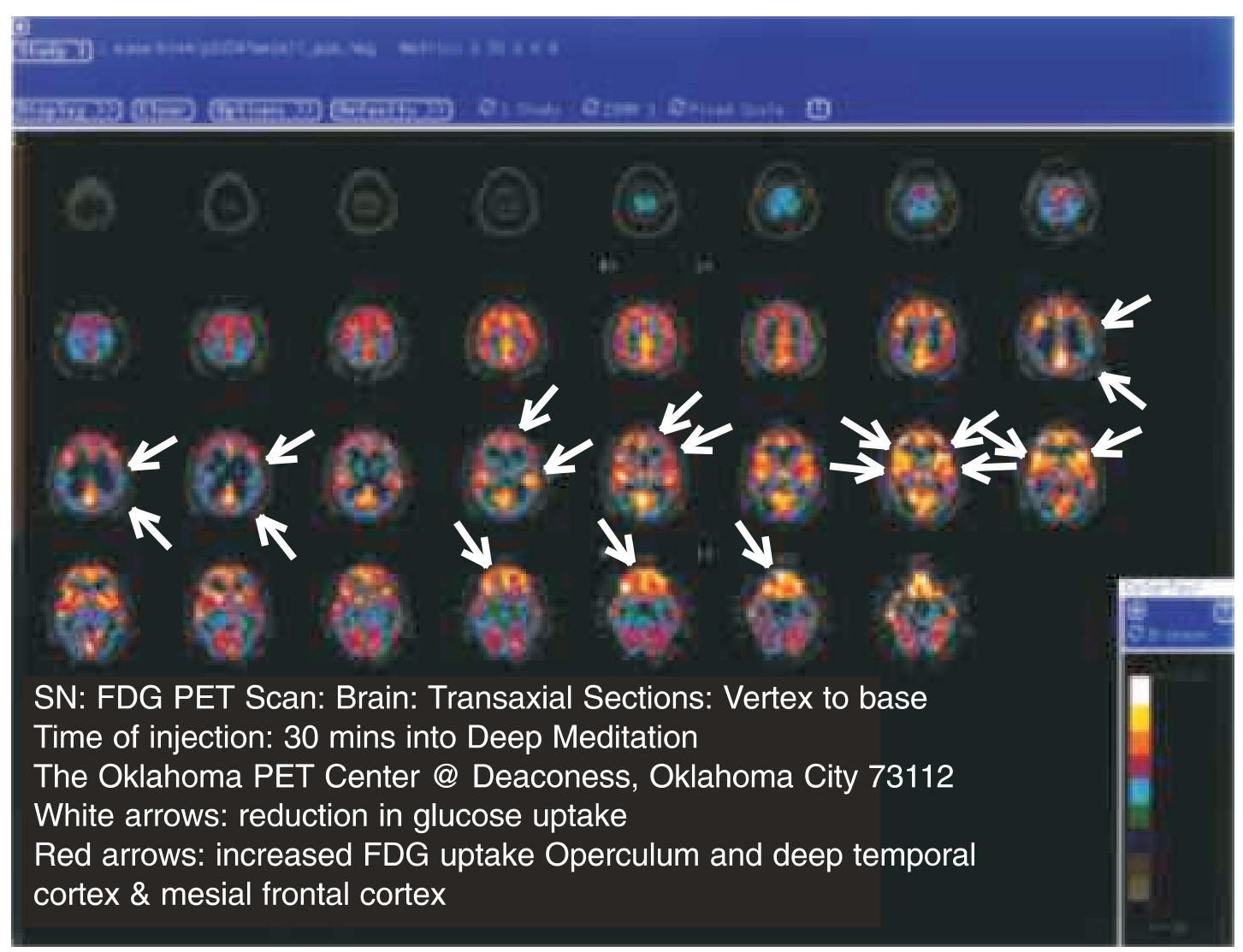
SN: FDG PET Scan: Brain: Transaxial Sections: Vertex to base Time of injection: 30 mins into deep meditation The Oklahoma PET Center @ Deaconess, Oklahoma City 73112 White arrows: reduction in glucose uptake. Red arrows: increased FDG uptake Operculum and deep temporal cortex & mesial frontal cortex.
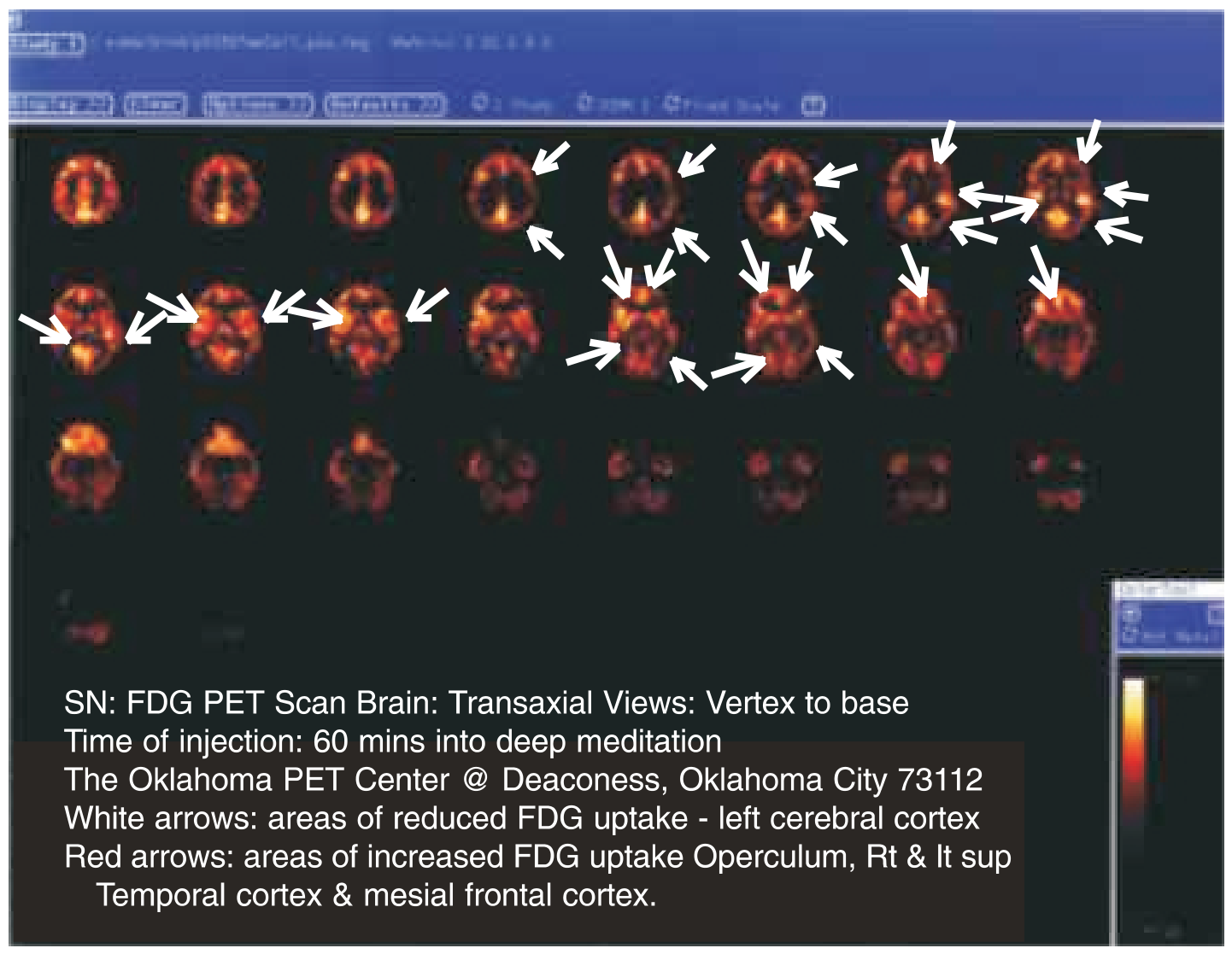
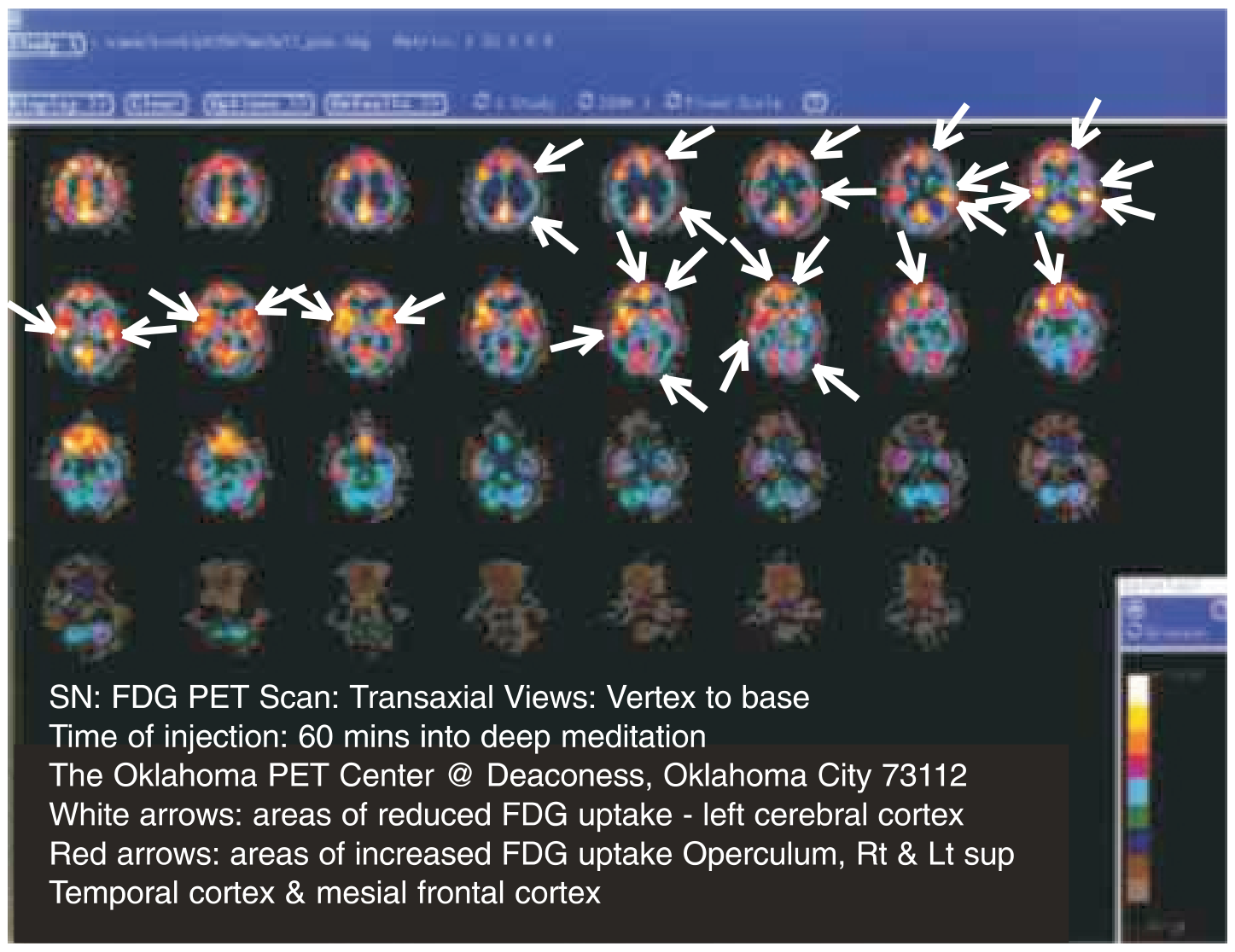
SN: FDG PET Scan Brain: Transaxial Views: Vertex to base Time of injection: 60 mins into deep meditation The Oklahoma PET Center @ Deaconess, Oklahoma City 73112 White arrows: areas of reduced FDG uptake - left cerebral cortex Red arrows: areas of increased FDG uptake Operculum, Rt & lt sup Temporal cortex & mesial frontal cortex.
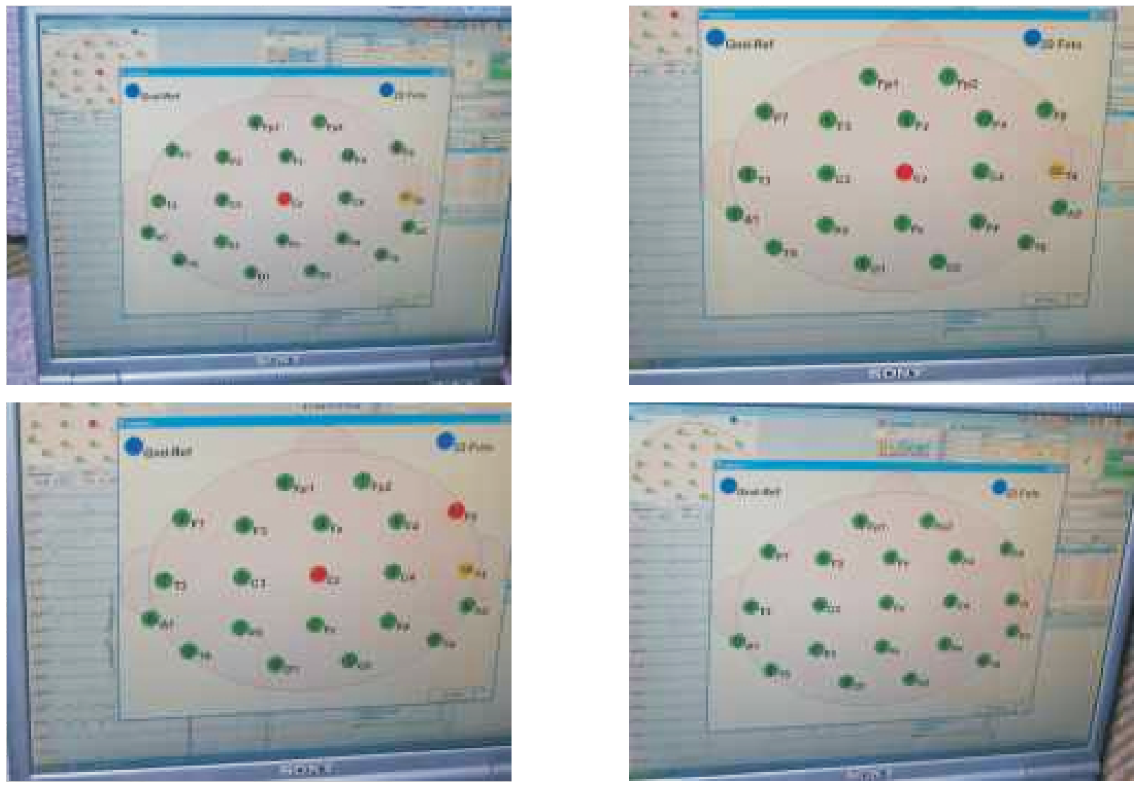
Pictures of electrode placement